











Urgent rib injuries seen same day · routine appointments within 24–48 hours 020 7459 4367 WhatsApp info@marcoscarci.co.uk
Chest wall lumps · swellings · masses · growths

A lump on the chest wall — the vast majority are benign, but all deserve a clear answer.

Finding a lump on the chest or ribcage is alarming. The good news is that most chest wall lumps are benign — lipomas, post-traumatic swellings, fracture callus or benign cartilage growths. A specialist assessment reaches the right diagnosis efficiently and avoids unnecessary anxiety or missed pathology.

Page last updated 5 August 2026

Most benignthe majority of chest wall lumps have a benign cause
Ultrasound firstquick, painless, no radiation — the usual starting point
Not breastbreast lumps need a breast clinic, not this service
Prompt biopsyonly when imaging cannot give a confident diagnosis
What it might be

Common causes of chest wall lumps — from the benign to the important

After trauma · most common

Post-traumatic swelling & haematoma

Following a chest wall injury, bruising and soft-tissue swelling are normal. A haematoma — a collection of blood in the chest wall tissue — develops in the hours to days after significant trauma. Most resolve on their own; large, tense or infected haematomas may need drainage. Rib fractures healing in poor alignment can produce a firm, palpable callus at the fracture site that persists for months.

Fatty tissue · very common

Lipoma

A benign tumour of fat tissue — soft, smooth, mobile, usually painless and slow-growing. Lipomas can occur anywhere on the chest and upper back. Most can be diagnosed clinically or with ultrasound without a biopsy. Removal is straightforward and only needed if the lump causes symptoms or significant concern.

Cartilage & bone · benign

Osteochondroma & bony exostoses

Benign bony outgrowths from the rib surface — often coincidentally discovered on imaging. Most cause no symptoms and need no treatment. Where a bony spur causes chronic friction, pain or restriction of movement, it can be trimmed surgically as a day procedure.

Bone · benign

Fibrous dysplasia

A benign bone disorder where normal bone is replaced by fibrous tissue — can cause rib pain, deformity or an incidental finding on a chest X-ray. Most monostotic (single rib) cases are asymptomatic. Symptomatic lesions with pathological fracture or deformity can be surgically stabilised.

Infection · treatable

Abscess & chest wall infection

A chest wall abscess presents as a hot, tender, fluctuant swelling — often developing after a skin infection, post-operative wound complication, or rib osteomyelitis (bone infection). Requires drainage and antibiotics; untreated empyema can occasionally extend into the chest wall from the pleural cavity.

Needs prompt review

Rib or sternal metastasis

Secondary cancers (particularly from breast, lung, thyroid, kidney or prostate) can spread to the bony chest wall, causing a painful, hard, often bony lump. These are among the more important diagnoses not to miss. A firm, painful bony lump in a patient with a cancer history — or an unexplained bony destruction on imaging — needs urgent specialist assessment.

This clinic does not treat breast lumps. Any lump that appears to be within breast tissue should be referred directly to a symptomatic breast clinic, which your GP can arrange as a two-week wait referral. The chest wall clinic assesses and treats lumps arising from the ribs, sternum, costal cartilage, intercostal muscles and other chest wall structures.

The diagnostic pathway

Reaching the right answer efficiently

Clinical assessment

A careful history — when it appeared, whether it is growing, associated symptoms — and a focused examination including palpation, percussion and auscultation. Many benign lumps can be confidently diagnosed at this stage alone.

Ultrasound

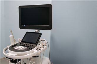
First-line imaging for most chest wall lumps: distinguishes fluid from solid, superficial from deep, and gives real-time dynamic information without radiation. Immediately available at the same hospital visit.

CT or MRI

CT provides excellent bony detail and is the standard for evaluating rib and sternal lesions. MRI characterises soft-tissue components and bone marrow involvement, particularly useful where the CT finding is indeterminate.

Biopsy (where needed)

Only performed when imaging cannot give a confident benign diagnosis. Core-needle biopsy under imaging guidance is safe, accurate and avoids open surgery for the diagnostic step. If malignancy is confirmed, management involves the oncological team.

Get an urgent assessment if the lump:

Growth

Is growing noticeably over weeks rather than months.

Texture

Is hard, fixed, or bony in character.

Pain

Is painful at rest or disturbs sleep.

General symptoms

Is associated with unexplained weight loss, fever or night sweats.

Cancer history

Appears in a patient with a known or previous cancer diagnosis.

Skin change

Has caused a visible deformity or skin change over it.

Neurological

Is associated with neurological symptoms (arm weakness, numbness).

Most urgent referrals can be seen within 48 hours. Where there is genuine clinical urgency, same-day assessment is available.

Patient outcomes

Three lumps, three answers — all of them fast

★★★★★

“I found a hard lump on my rib in the shower and spent two weeks convincing myself of the worst. Mr Scarci saw me within days, examined it, and the CT showed a benign bony outgrowth I’d probably had since my teens. He explained every image on the screen. I cried with relief in the car park. We removed it a month later purely because it caught on my bra strap.”

Benign rib osteochondroma, excised · verified review
★★★★★

“A swelling came up on my chest a week after a heavy gym session and Google had me terrified. The ultrasound happened right there in the consultation — a haematoma from a strained muscle, nothing more. Diagnosed, explained and reassured inside forty minutes. It was gone on its own within six weeks, exactly as he predicted.”

Post-injury haematoma, reassured same day · verified review
★★★★★

“Mine was the rare kind. Mr Scarci never dressed it up — he told me straight, organised the biopsy that week, and had me in front of the specialist team within days with everything already staged and prepared. If it had to be bad news, this was the way to receive it: fast, honest, and never once feeling alone in it.”

Chest wall tumour, specialist pathway · verified review

Read 135+ verified reviews

Mr Marco Scarci, consultant thoracic surgeon
Your surgeon

Mr Marco Scarci

MD(Hons) · FRCS(Eng) · FCCP · FACS · FEBTS · Consultant Thoracic Surgeon, Imperial College Healthcare NHS Trust

Chest wall lumps sit squarely in thoracic surgical territory: the ribs, sternum, cartilage and their soft tissues are the structures Mr Scarci operates on, images and biopsies as his core work — including, in his NHS practice, the full spectrum from benign outgrowths to chest wall tumours. That breadth is what a lump assessment needs most: the experience to recognise the harmless confidently, the discipline to image everything anyway, and the judgement to move fast on the rare lump that demands it.

Every examination is performed by Mr Scarci personally, every scan reviewed by him with you at the screen, and every result — reassuring or not — delivered by him in person.

20+years as a consultant thoracic surgeon
5,000+minimally invasive thoracic procedures
170+peer-reviewed publications
Read the full biography
Book an assessment

Seen the same week — five London clinic locations

No GP referral required. If you already have scans or reports, bring them — and if the lump has been photographed over time, those photos are genuinely useful evidence.

Not in London? Remote video consultations are available to review your history, photographs and any existing imaging before you travel.

BupaAXA HealthAviva VitalityCignaWPA HealixSelf-pay · 0% finance
Common questions

Chest wall lumps — what patients ask

Should I be worried about a lump on my chest wall?

Most chest wall lumps are benign — the vast majority turn out to be lipomas (fatty lumps), fracture callus (bone healing), haematomas (post-injury blood collections), or benign cartilage and bony growths. However, some do require treatment and a small minority are malignant, so any unexplained lump that is new, growing, hard, painful, or associated with other symptoms warrants expert review. The purpose of a specialist consultation is to reach the right answer efficiently — not to alarm you.

What investigations will I need?

This depends entirely on the lump's character. Ultrasound is usually the first step: quick, painless, no radiation, and excellent for distinguishing fluid-filled, fatty and solid lumps. CT or MRI is used for deeper or bony lesions, or where malignancy needs to be properly excluded. A biopsy is only performed when imaging cannot give a confident benign diagnosis — not routinely. Many lumps are diagnosed clinically and need no further testing at all.

Can a chest wall lump be cancerous?

The majority are not. Primary malignant chest wall tumours (sarcomas) are rare. Secondary metastases from breast, lung, thyroid or other cancers can spread to the chest wall and ribs, and these are more common than primary chest wall cancers. Painful bony lumps with no clear traumatic cause, lumps that are growing rapidly, or any lump in a patient with a known cancer history should be assessed promptly. Where malignancy is confirmed, the management involves the wider oncological team, with Mr Scarci providing the thoracic surgical component.

I this clinic the right place for a breast lump?

No. This clinic does not assess or treat breast lumps — any concern about a breast lump should go directly to a breast clinic or via your GP to a symptomatic breast service. The chest wall clinic handles lumps arising from the chest wall structures themselves: ribs, costal cartilage, sternum, intercostal muscles, subcutaneous tissue and the chest cavity lining.

How are benign chest wall lumps treated?

Many do not need treatment at all — a firm diagnosis, reassurance, and watchful waiting are entirely appropriate for stable benign lesions such as small lipomas or incidental fibrous dysplasia. Where a lump causes pain, restricts movement, is growing, or causes anxiety despite clinical reassurance, surgical excision is usually straightforward. Post-traumatic haematomas and abscesses may need drainage. Bony exostoses (spurs) causing chronic friction or pain can be trimmed surgically.

A chest wall lump needs a confident diagnosis — reassurance or treatment, whichever is right.

One consultation with clinical examination, immediate imaging and a clear plan. Same-day assessment available for urgent cases.