Chest wall pain after injury — when the cartilage is the problem, not the bone.
The chest wall is more than ribs and sternum. Every rib connects to the breastbone
via a costal cartilage — and the joints between bone and cartilage are often injured in exactly the
same incidents that fracture ribs. Crucially, they are largely invisible on X-ray. Thousands of
patients are discharged with “normal” imaging when the cartilage junction is the actual source
of their pain.
X-ray invisiblecostal cartilage does not show on plain films
12 junctionssix ribs each side with distinct cartilage connections
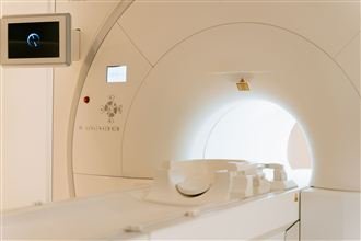
CT or MRIneeded to image cartilage injuries properly
Often mistreatedmanaged as muscle pain or anxiety
The anatomy you need to know
Four junctions — four different injury patterns
Each cartilage junction in the chest has its own mechanics and its own characteristic injury pattern.
Understanding which junction is affected is the key to getting the right treatment.
Top of sternum
Manubriosternal joint
Where the manubrium (upper sternum) meets the body of the sternum at the sternal angle — felt as
a natural ridge in the middle of the chest. High-energy trauma (seatbelt, steering wheel, RTA) can
dislocate this joint, causing severe central chest pain and visible or palpable deformity. Lower-energy
injuries produce localised inflammation and pain. CT is needed to characterise the injury.
Front of chest · ribs 1–7
Costo-sternal junction
Where the cartilage of the upper ribs attaches to the sternum. A direct blow, indirect
deceleration or even a violent cough can sprain, sublux or fracture this junction. The pain is
immediate and very localised — tender to press, worse with breathing and movement. Tietze syndrome
(inflammatory, with visible swelling) and simple costochondritis (inflammatory, no swelling) both
affect this junction.
Lateral · away from sternum
Costochondral junction
Where the bony rib meets its costal cartilage further out from the breastbone. Injuring this
junction produces point tenderness lateral to the sternum — a location that surprises many patients
who expect chest injuries to hurt in the middle. Subluxation gives a palpable click or step. This
junction is particularly vulnerable in sports with twisting or impact.
Lower ribcage · ribs 7–10
Costal arch & interchondral junctions
The costal arch is formed by the costal cartilages of ribs 7–10 meeting at the lower chest margin.
The interchondral junctions between these cartilages can sublux or dislocate, causing a lower-rib
pain that overlaps with slipping rib syndrome. Costal arch injuries are the most mechanically complex
and hardest to image — dynamic ultrasound is often the most useful investigation.
The xiphoid process at the very tip of the breastbone can become inflamed or fractured, producing
epigastric pain that mimics gastric or cardiac causes. Pressure or bending aggravate it. Treatment
ranges from reassurance and anti-inflammatories to local injection and, in persistent confirmed
cases, surgical xiphoidectomy.
Both cause pain at the costo-sternal junctions without a traumatic cause. Tietze syndrome is
distinguished by visible, palpable cartilage swelling (usually rib 2 or 3); costochondritis is more
widespread with no swelling. Most settle with anti-inflammatories; persistent or severe cases respond
to local steroid injection.
Getting the right diagnosis
Why junction injuries are so often missed
Three things conspire to leave cartilage injuries undiagnosed for months or years:
X-rays look normal
Cartilage is radiolucent; fractures and subluxations at these junctions simply do not show on plain films.
CT is often not requested
After a chest injury, CT is typically ordered to look for pneumothorax and haemothorax, not to image cartilage junctions specifically.
No specialist examination
The specific palpation tests for each junction are not standard in ED or orthopaedic assessment, so the diagnosis is never even considered.
The result is that patients are typically told their ribs are fine, given analgesia, and sent home
— then left with months of unexplained pain. The assessment here starts with a clinical
examination specifically designed for the chest wall junctions, and uses CT or MRI specifically
targeted at cartilage when needed.
A dedicated fine-slice CT with 3D reconstruction of the costal cartilage shows fractures, dislocations and structural abnormalities at each junction that plain X-ray cannot see.
Soft tissue & inflammation
MRI
Shows bone marrow oedema (a sign of acute injury), cartilage integrity and inflammation around the affected junction — particularly useful for Tietze syndrome and manubriosternal injuries.
Dynamic
Ultrasound
Can image the lower costal cartilages in real time as the patient moves — critical for demonstrating interchondral subluxation and costal arch instability that static imaging misses.
Diagnostic & therapeutic
Local steroid injection
Injecting corticosteroid directly into the affected junction reduces inflammation and confirms the location by providing immediate relief — often performed under ultrasound guidance for accuracy.
For persistent instability
Cartilage excision / stabilisation
When a costal arch or interchondral junction remains unstable and painful despite conservative measures, surgical excision of the unstable cartilage tip resolves the mechanical cause of the pain.
Patient outcomes
Patients whose “normal” scans were hiding a real injury
“★★★★★
“I felt something tear on my downswing and the pain was instant. A&E X-rayed me, found nothing, and said bruising. Four months later I still couldn’t play. Mr Scarci found the exact junction in about a minute, and the ultrasound showed the separation moving on screen — there it was, the injury everyone said didn’t exist. An injection and a proper graded return, and I was back on the course by autumn.”
“★★★★★
“A brutal chest infection left me with a click at my lower ribs every time I twisted or laughed. Eight months of being told it was muscular — one GP suggested anxiety. Mr Scarci examined the rib arch, diagnosed a costal margin rupture from the coughing, and repaired it surgically. The clicking stopped the day of the operation and has never returned.”
“★★★★★
“Something popped at the top of a heavy bench press and every press after that was agony next to my breastbone. I was told costochondritis and to give it time — for a year. The MRI Mr Scarci organised showed the damaged cartilage segment, and after excision I’m training pain-free for the first time since the injury. A year of waiting for the wrong diagnosis to heal.”
Cartilage junction injuries fall through a gap in the system: emergency medicine excludes the fracture, sports medicine treats the muscles, and the cartilage in between belongs to nobody — except the thoracic surgeon, whose entire operating field is the bone-and-cartilage architecture of the chest wall. Mr Scarci has spent over two decades in that territory: examining the junctions others never palpate, choosing imaging that can actually render cartilage, and performing the repairs — margin stabilisation, cartilage excision, slipping rib resection — that resolve the injuries which refuse to heal.
Every examination, injection and repair is performed by Mr Scarci personally, from first consultation to final review.
Because cartilage is largely invisible on plain X-rays. The bony ribs and sternum show clearly; the costal cartilage connecting them does not. A costochondral fracture, costo-sternal subluxation or costal arch injury can be completely invisible on X-ray but very visible on CT or MRI, and definitively confirmed by clinical examination. A normal X-ray after a chest injury does not mean nothing is wrong with the cartilage.
What does a costochondral injury feel like?
Sharp pain exactly at the point where the rib meets its cartilage — typically felt on the front of the chest, away from the breastbone. It is usually exquisitely tender to press. Deep breaths, twisting and certain positions make it much worse. In cases with subluxation (partial dislocation) there may be a palpable click or step that can be felt. Unlike rib fractures, which tend to hurt along the whole rib, costochondral pain is very localised.
How long does a costochondral injury take to heal?
Simple costochondral sprains usually settle within 6–12 weeks with appropriate pain management and activity restriction. Displaced or subluxed junctions may take longer and can cause chronic pain if the instability is not addressed. Manubriosternal injuries from high-energy trauma sometimes require surgical stabilisation if significantly displaced. Costal arch subluxation overlaps with slipping rib syndrome and may need cartilage excision for lasting resolution.
Is there a difference between Tietze syndrome and a costochondral injury?
Tietze syndrome is an inflammatory condition affecting the same junction (costo-sternal), typically presenting with pain and visible or palpable swelling at the second or third rib. Simple costochondritis produces identical pain but without swelling. Traumatic costochondral injuries are caused by a specific force and often involve structural disruption rather than pure inflammation. All three can present similarly and require examination to distinguish them, which is exactly why a targeted chest wall assessment is more useful than another scan.
What treatments help junction injuries?
Initial treatment is always conservative: relative rest, regular anti-inflammatories, avoidance of aggravating movements, and targeted physiotherapy to maintain mobility without overloading the injured junction. Where pain is persistent, local steroid injections into the affected joint can provide significant medium-term relief. For costal arch injuries that have become mechanically unstable (effectively slipping rib syndrome at the costal margin), surgical cartilage excision is the definitive solution.
When it is an emergency — call 999
Sudden severe breathlessness at rest, coughing up blood, blue lips or fingertips, crushing central chest
pain, fainting, or a section of the chest visibly moving the wrong way with breathing: these need an
emergency ambulance, not an outpatient appointment.
Chest wall pain after injury with a normal X-ray is not normal — it just means the right structure hasn't been imaged yet.
A dedicated chest wall assessment with targeted imaging and examination of every junction usually finds the answer within one visit.
This site uses essential cookies to work correctly. With your consent we'd also like to use analytics cookies to understand site usage — see our Cookie Policy for details.