
Clinical examination
History plus a positive hooking manoeuvre is sufficient to make the diagnosis — and in published series the examination findings closely match what is found at surgery.
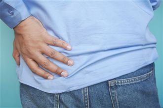 Slipping rib syndrome · Cyriax syndrome · clicking rib
Slipping rib syndrome is one of the most consistently missed diagnoses in medicine: sharp pain under the lower ribs, sometimes a click or pop, and test after test reported as normal. It is real, it has a name, and it is diagnosed in minutes by a clinician who knows exactly what to look for.
Page last updated 5 August 2026











It is the sentence patients say most often at their first appointment — years of pain, and every scan reported as normal. Slipping rib syndrome does not show up on standard imaging; the diagnosis is made by examination, not by scans.
“The tragedy of slipping rib syndrome is not the condition — it is the years patients spend being investigated for everything else before anyone performs a proper rib examination. One clinical test often makes the diagnosis that five years of scans could not.”
Mr Marco Scarci, Consultant Thoracic Surgeon
It is regularly mistaken for: gallbladder disease (in one published series roughly one in five patients had already had their gallbladder removed — and their pain continued), costochondritis, reflux and IBS, intercostal neuralgia, muscle strain, cardiac pain, and — once everything else is excluded — “functional” or stress-related pain.
Most affected patients in published series are women, and many were told their symptoms were stress-related before the correct diagnosis was made.
Swimmers, rowers, golfers, tennis players, gymnasts — repetitive trunk rotation gradually strains the ligaments between the lower ribs.
Generalised ligament laxity makes rib instability far more likely, often at several ribs — and changes how any surgery should be planned.
Hormonal ligament changes plus the physical load of carrying and lifting a baby can unmask rib instability, sometimes months after delivery.
Car accidents, falls, direct blows — even violent coughing fits — can tear the ligaments that keep the false ribs stable, with symptoms appearing weeks later.
Sustained lifting, twisting and overhead reaching load the costal margin day after day, gradually loosening its attachments.
A recognised minority of patients date their symptoms to laparoscopic surgery — gallbladder, appendix or gynaecological procedures.
A deliberately simple bedside examination that reproduces the slip itself — confirming the diagnosis and identifying exactly which rib is responsible.
You lie back comfortably while the examiner feels along the lower rib margin for the point of maximum tenderness.
The examiner curls their fingers gently beneath the rib edge and applies careful upward and outward traction.
If the rib is unstable, this reproduces your familiar pain — often with the characteristic click — felt by you and the examiner.
The other side is checked too; the rib(s) that test positive guide the treatment plan.
History plus a positive hooking manoeuvre is sufficient to make the diagnosis — and in published series the examination findings closely match what is found at surgery.
Ultrasound performed while the rib is actually moving can visualise the slip directly — useful confirmation in selected cases, in experienced hands.
Local anaesthetic beside the affected nerve that abolishes the pain both confirms the source and gives relief — invaluable for surgical planning.
Useful for ruling other things out — but they cannot see the dynamic laxity of a slipping rib. A normal scan does not close the question.
Once the diagnosis is confirmed, physiotherapy addresses breathing mechanics, core stability and the compensation patterns that let the rib slip — supported by simple analgesia and lidocaine patches.
Local anaesthetic — with corticosteroid where appropriate — alongside the affected nerve. Days to weeks of significant relief for many patients, and precise confirmation of which rib is responsible.
For patients whose quality of life remains severely limited: the unstable cartilage tip is removed through a small incision, eliminating the mechanical cause of the pain. Combined where appropriate with costal margin stabilisation (vertical plating), a rib-preserving sutured repair in selected cases, or intercostal nerve cryoablation for long-sensitised nerves.
★★★★★“I had rib pain for six years and bounced around the NHS without a diagnosis. Within minutes he knew what the problem was — slipping rib syndrome. Surgery the following week. Two weeks later I’m almost back to full health. Simply amazing.”
★★★★★“I’d stopped going to yoga, avoided hugging my children, and couldn’t sleep on my left side. Every time it slipped I felt sick with the pain. After surgery I could take a full breath without bracing for something to go wrong. It changed everything.”
★★★★★“I’d seen five specialists over three years. Every scan was normal. I’d been told it was stress, then a pulled muscle. Mr Scarci did the hooking test in the first five minutes and said ‘I know exactly what this is.’ I cried with relief.”

MD(Hons) · FRCS(Eng) · FCCP · FACS · FEBTS · Consultant Thoracic Surgeon, Imperial College Healthcare NHS Trust
Slipping rib syndrome sits in a gap between specialities — too structural for physicians, too specialised for general surgeons, and invisible to imaging. Mr Scarci has spent over two decades building the specific clinical skill to identify and treat it: the bedside examination technique, the surgical approach for costal cartilage excision, and the modifications needed when hypermobility is in the picture.
He has personally performed the hooking manoeuvre on hundreds of patients with long diagnostic delays, and operates on the select cases where surgery is the right answer. He sees you personally at consultation, performs any surgery himself, and reviews your recovery.
Choose the day and hospital that suits you. The hooking manoeuvre will be performed at your first visit.
St John’s Wood, NW8 9LE
11:00am–1:00pm Monday PMMarylebone, W1G 6BW
5:30–7:30pm Tuesday PMBorehamwood, WD6 3BS
5:30–7:30pm Wednesday AMHammersmith, W12 0HS
9:00–11:00am Thursday AMSouth Kensington, SW5 0TU
9:30am–12:30pmNot in London? Remote video consultations are available for review of existing imaging and initial assessment before you travel.
The classic pattern: sharp pain under the lower ribs, usually one-sided, triggered by specific movements — twisting, bending, reaching, rolling over in bed — often with a clicking or popping sensation, and with normal scans despite very real pain. The definitive check is a clinical examination including the hooking manoeuvre. If no one has ever performed that test on you, the diagnosis has never actually been assessed.
Because the problem is mechanical and movement-dependent. X-ray, CT and MRI photograph the rib at rest, when it sits in a normal position; the abnormal movement — the slip — only happens when you move. In published series the great majority of confirmed patients had prior imaging, and none of it made the diagnosis. A normal scan does not mean nothing is wrong; it means the problem is not visible at rest.
A simple bedside test: the examiner curls their fingers under the lower rib margin and applies gentle upward and outward traction. If the rib is unstable, this reproduces your familiar pain — often with the click — confirming both the diagnosis and exactly which rib is responsible. In published series the examination findings closely match what is found at surgery, which is why no scan can substitute for being properly examined.
Often, yes. Targeted physiotherapy, activity modification and intercostal nerve blocks control symptoms well for many patients, particularly milder cases. But the underlying problem is structural ligament laxity — conservative care manages the consequences rather than the cause. For severe, frequent episodes that dominate daily life, surgery is the only treatment that addresses the mechanics directly.
Costal cartilage excision removes the unstable cartilage tip — most commonly rib 8, 9 or 10 — through a small incision of a few centimetres at the lower rib margin, under general anaesthetic. Depending on your anatomy, this is combined with stabilisation (for example vertical plating of the costal margin) or, in selected patients, a rib-preserving sutured repair. Nerve cryoablation can be added where years of irritation have sensitised the intercostal nerve. Hospital stay is typically one to three nights, with a return to light activity in two to four weeks.
Yes, meaningfully. Generalised ligament laxity makes rib instability more likely, more often affects several ribs, and raises the recurrence risk after simple cartilage excision alone — so stabilisation techniques and carefully staged rehabilitation matter more. It is worth being treated by someone who regularly manages the combination of hypermobility and chest wall mechanics.
Consultations, diagnostic nerve blocks and surgery for slipping rib syndrome are recognised by the major UK insurers as a chest wall condition, and the practice handles authorisation for you. Self-pay patients receive transparent written estimates, and remote video consultations are available if you are not in London — many patients travel for this diagnosis.
One consultation with a specialist who understands chest wall mechanics can give you the answer years of normal scans could not — and a clear plan for what to do about it.